What is Imaging in Dental Treatments?
In dental treatments, imaging plays a critical role in diagnosing problems in the mouth and jaw structure (cavities, impacted teeth, bone loss) with details that are not visible to the naked eye. Thanks to digital methods in modern dentistry, both radiation is minimized and diagnostic accuracy is increased.
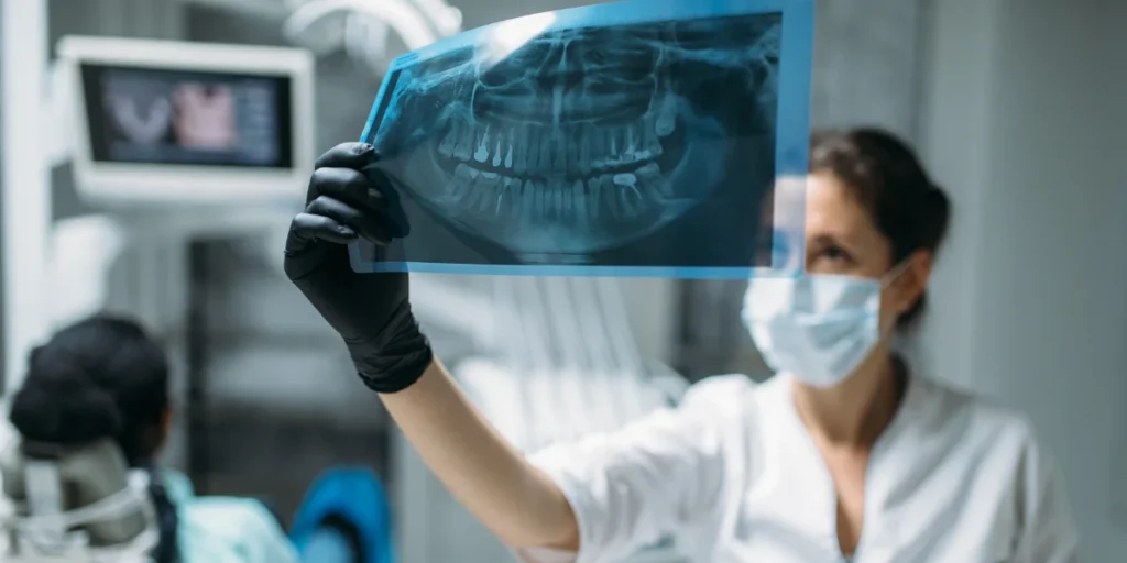
Panoramic X-ray: Decayed and impacted teeth, which show the overall structure of the jaw and teeth, are the first choice for procedures such as implants before orthodontics (braces).
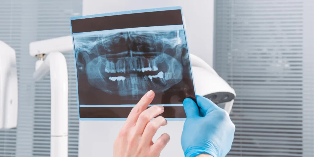
Reasons for Obtaining a Panoramic X-ray
General Oral Health Check: During the initial dental examination, a panoramic view of the jaw structure is taken.
Tooth Development Monitoring: In children, the direction of eruption of permanent teeth, the condition of primary teeth, and jaw development are monitored.
Treatment planning: It is used in many procedures for general assessment before dentures, implants, and braces.
3D Tomography (3D):
It provides a three-dimensional and more detailed view of the jaw. The volume of the jawbone, the roots and location of the nerve canals are checked. In impacted tooth surgeries, whether the teeth are close to the bone or the position of implants or cyst surgeries is evaluated.
Areas of Application
- Implant treatment: Bone density and suitability for implants are checked.
- Root Canal Treatment: Determining the structure of the root canals.
- Impacted Teeth: The position of teeth and their relationship with surrounding tissues.
- Temporomandibular Joint (TMJ): Examination of the joint structure.
- Root Infections: Inflammation or pathological conditions are detected in the tooth roots.
- Cyst and Tumor Diagnosis: Hidden infections, fractures, cracks, and joint problems in the jawbone that cannot be detected during examination are investigated.
- Orthodontics: Analysis of the teeth and jaw structure.
Panoramic X-ray Imaging in Children
In children over 6 years of age, X-rays play a crucial role in assessing oral development and detecting cavities that are not visible to the naked eye. In younger children, X-rays are taken after an oral examination if deemed necessary.
- Periapical X-ray: It is generally used in endodontics (root canal treatment). It shows only a few teeth and their root tips in detail. It is used for checking the roots during and after treatment, and for teeth with local inflammation.
- Digital Intraoral Scanning (3D Scanning): It is generally used with high precision in denture and clear aligner treatments. It is a radiation-free method that creates digital models of teeth, replacing clay impressions.
For pricing information regarding imaging procedures, please contact İnci Diş.