
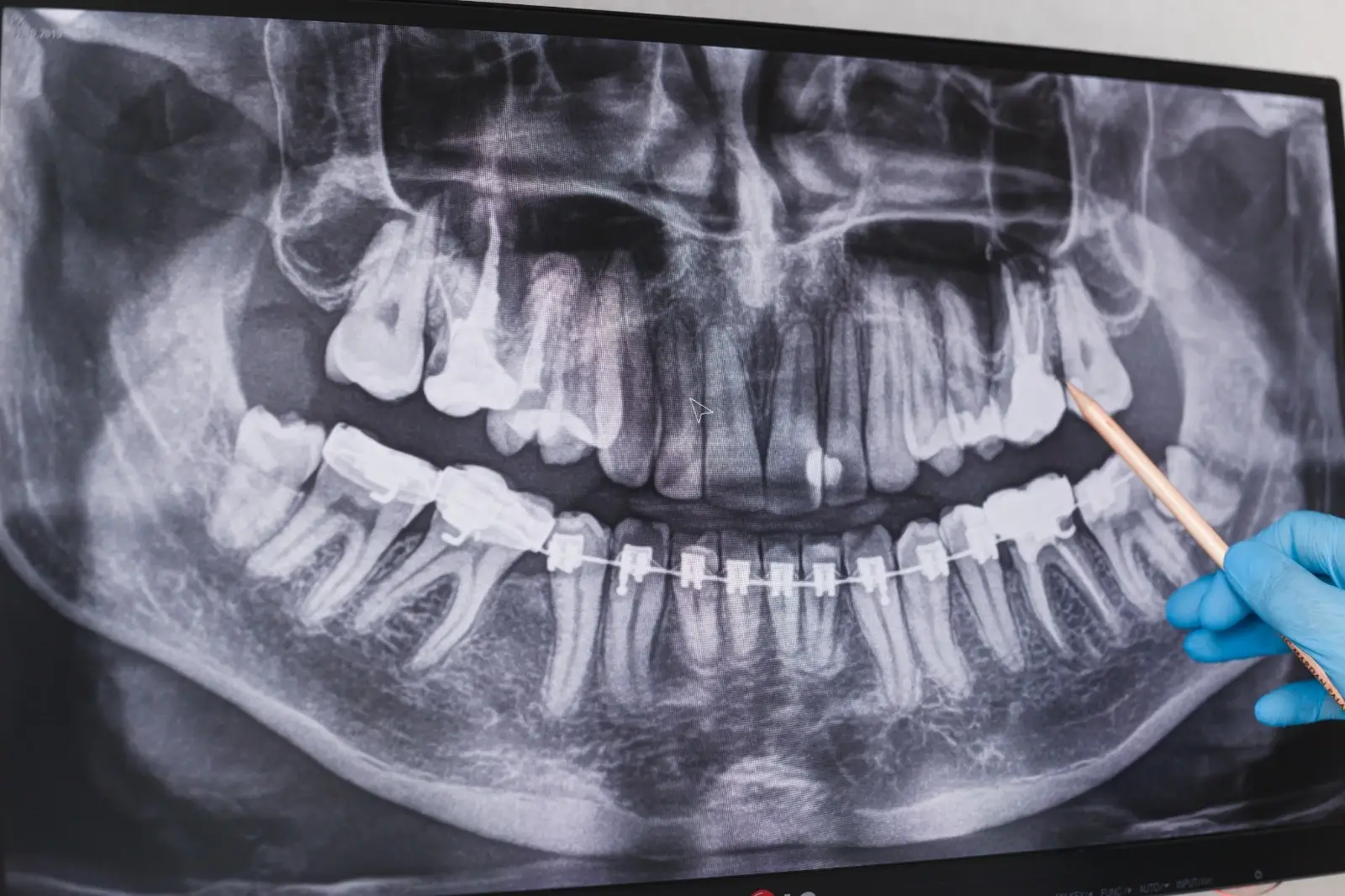
Dental X-ray
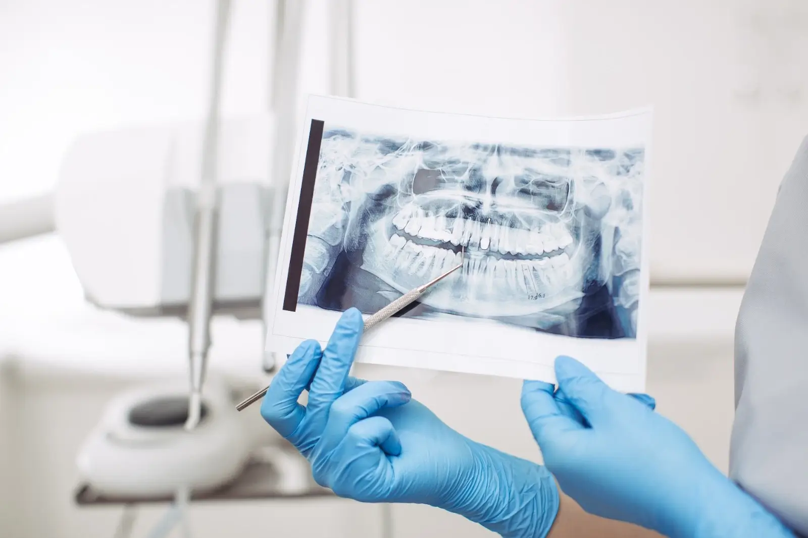
Dental X-rays, or radiographs, are radiographic images used by dentists to clearly visualize patients' oral health and the condition of tooth roots. Taking an X-ray before a dental examination is essential for a detailed examination of the mouth, jaw, and tooth structure. The dentist can see images of decayed teeth, spaces between teeth, and tooth positions. This plays a crucial role in making an accurate diagnosis and applying the correct treatment during the examination.
Contents
- Why are dental X-rays taken?
- Are there any disadvantages to dental X-rays?
- What should be done before a dental X-ray?
- What are the different types of X-rays used in dentistry?
- Are X-rays Harmful to Children?
- How often is it appropriate to have X-rays?
- What are the advantages of dental X-rays?
- Fill out the form to get detailed information.
Why are dental X-rays taken?
Dental x-ray, oral examination A prior examination is performed to provide a clearer and more detailed view of any problems in the teeth. Sometimes, it may not be possible to detect problems in the tooth roots during an oral examination, or the patient's jawbone (jawbone pain If there is a problem related to (etc.), it is quite difficult to detect this problem without seeing an X-ray. X-rays are of great importance in jaw and dental treatments. X-ray image of a decayed tooth., image of an impacted tooth, implant Problems such as bone adequacy or lesions that may occur in the root can only be detected by X-ray.
Are there any disadvantages to dental X-rays?
Dental X-rays contain minimal radiation. Therefore, they pose no danger to children or adult patients. During the X-ray, the patient wears a protective lead apron over their chest. It is especially important for child patients to wear this apron. However, X-rays pose a risk to pregnant women due to the radiation they contain, and exposure to this radiation is not advisable. Even if pregnancy is suspected, this must be mentioned during the medical history.
What should be done before a dental X-ray?
No specific preparation is needed for an X-ray. The X-ray is taken in the dental clinic with the patient wearing a protective lead apron on their chest. The X-ray machine is positioned around the patient's skull and rotates to record images of the inside of the mouth. Clinics have a separate X-ray area. The walls and door of this area must be lead-lined to prevent radiation from spreading to the surrounding area.
What are the different types of X-rays used in dentistry?
Generally, dental X-rays are divided into four types: periapical X-ray (showing a single tooth), standard panoramic X-ray, cephalometric X-ray, and dental tomography. Let's examine these further.;
- Periapical X-ray
This type of X-ray provides a complete and clear image of the tooth, including the roots, and is therefore a very frequently used type of X-ray. Periapical X-rays are preferred when the root structure and the condition of the tooth in relation to the bone need to be evaluated. They play an important role in detecting various conditions such as cysts and abscesses, as well as the tooth roots and bone level. They are most commonly used in endodontics. Root canal specialists frequently use periapical X-rays to examine the tooth in detail. Periapical X-rays are taken from only one area; they do not show the entire mouth.
- Standard Panoramic Dental X-ray
Often, imaging the entire oral cavity is necessary to identify the factors causing complaints such as pain or abscesses in patients. This is where the standard panoramic dental X-ray comes into play. This type of X-ray allows for the examination of all teeth in the upper and lower jaws, as well as a portion of the jawbone, in a single image. Panoramic dental X-rays are also used to examine the development of both primary and permanent teeth in children, in addition to adults. When patients first come for an examination, a general assessment is necessary. Based on this, a standard panoramic X-ray is taken.
- Cephalometric Dental X-ray
Cephalometric dental X-rays are a radiographic examination that provides visualization of the skull bones and soft tissues in anterior, posterior, and lateral positions. They may be needed for orthodontic treatments (braces) or in cases requiring surgery.
- Dental CT Scan
Dental CT scans are the preferred type of imaging to provide clear visualization of structures in a targeted treatment area. X-rays are used to perform dental CT scans, which consist of thin sections that also include volume. Although often compared to standard dental X-rays, there are significant differences between them. The biggest difference is that CT scans provide a 3D image. Therefore, it is possible to obtain findings such as length, depth, and width in the targeted area for treatment. It is a type of X-ray that provides visualization of all teeth, bone level, and jaw structure.
Are X-rays Harmful to Children?
The answer to whether panoramic radiography poses any danger to children depends on why it is recommended. Whether for dental or medical reasons, all types of radiography should only be performed when there is a clear need for the information they provide.
If children have clinically diagnosed dental problems (large gaps between teeth, swelling in the mouth, severe pain, or abnormal jaw development, etc.), a panoramic X-ray may be a good option and considered reliable in terms of determining the need for treatment. However, if the dentist does not detect any clinical problems and the child has no apparent symptoms, most current dental authorities do not recommend X-rays for very young children. If your child's dentist or your own dentist deems an X-ray necessary, it's a good idea to inquire about the reason.
How often is it appropriate to have X-rays?
The frequency of dental X-rays is related to the patient's oral and dental health and the risk of caries progression. While every one to two years is considered appropriate for adult patients, some patients with a high number of cavities and poor oral health may require X-rays every six months. There is a reason why bite radiographs, taken to check the spaces between teeth, are repeated periodically: clinical examinations cannot maintain continuity until the teeth become very large and difficult to treat. The frequency of X-rays depends on the patient's oral health and risk of developing cavities.
What are the advantages of dental X-rays?
Dental X-rays, or panoramic radiography as it's called in medical terminology, offer many advantages. Firstly, radiographs taken to determine the starting points of cavities in teeth provide the opportunity for more accurate treatment. Secondly, they allow for the visual detection of problems such as cysts or tumors caused by inflammation in the jaws, enabling the necessary and correct treatment. In this way, patients achieve the desired oral health.
Furthermore, one of the most important advantages of X-rays is the detection of problems in the tooth roots and jawbone that cannot be detected during an oral examination. Early diagnosis allows for all treatments to be performed promptly and before the problem worsens, resulting in a high success rate. Timely and correct intervention prevents post-treatment problems, avoids complex treatment methods, and saves both time and money. Dental X-rays have many advantages like these and have no disadvantages…
For further questions and answers, you can contact İnci Oral and Dental Health Centers and take advantage of our free examination service. Oral and Dental Health Center Inci Dental, which has become a leading brand in its sector and aims to add new achievements to its successes every day, offers free radiographic (X-ray) imaging and intraoral examinations in all its clinics.